

Executive Overview
To the untrained eye, any small bump on the facial landscape is instantly categorized as a blemish. Whether it is a rebellious whitehead, a stubborn blackhead, or a deep-seated cystic breakout, skin irregularities are a universal human experience. However, beneath the umbrella of everyday blemishes lies a deceptive impostor that masquerades as acne while operating under an entirely different biological rulebook: milia.
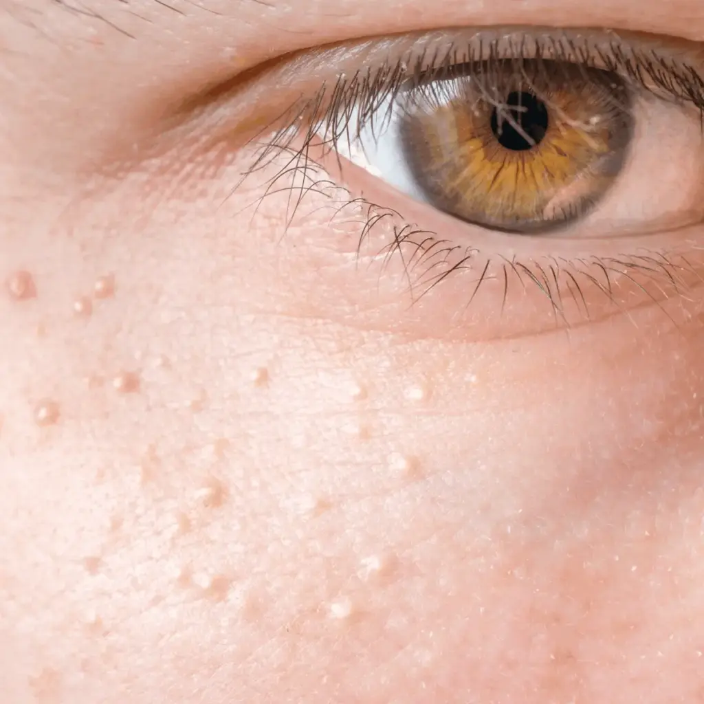
Characterized by tiny, pearlescent white dots that frequently cluster around the delicate eye contours, eyelids, cheeks, and forehead, milia are frequently misunderstood. While they bear a striking visual resemblance to closed comedones—the medical term for whiteheads—milia are not a form of acne at all. They do not stem from overactive sebaceous glands, bacterial proliferation, or trapped sebum within an open pore. Instead, they are miniature, benign keratin cysts trapped beneath the topmost layers of the epidermis.
Because these tiny cysts lack a natural exit route to the skin’s surface, treating them requires an entirely different approach than standard spot treatments or salicylic acid face washes. Attempting to manage them with conventional extraction methods often results in skin trauma, inflammation, and lasting scarring.
This comprehensive investigation explores the biological mechanisms behind milia, examines the primary triggers identified by leading dermatologists, separates at-home management myths from clinical realities, and outlines expert-backed prevention strategies to restore a smooth, flawless complexion.
Detailed Chronology: How Milia Form and Evolve Over Time
Understanding milia requires a journey into the microscopic architecture of human skin. To appreciate why these tiny bumps are so notoriously resilient, one must trace their lifecycle from initial cellular entrapment to their eventual resolution—whether natural or clinical.
Phase 1: The Cellular Trapping Mechanism
In a healthy, normally functioning integumentary system, skin cells undergo a continuous cycle of generation, migration, and shedding known as cellular turnover. New cells are produced in the basal layer of the epidermis, slowly making their way upward toward the stratum corneum (the outermost layer), where they naturally slough off, revealing fresh skin beneath.
During this process, cells produce a fibrous, protective structural protein called keratin. In cases where milia develop, this natural shedding process is temporarily interrupted. Instead of reaching the surface and dispersing into the environment, dead skin cells and excess keratin become trapped in a pocket just beneath the epidermis. As more protein accumulates in this microscopic pocket, it compacts into a firm, hard, spherical ball, forming a tiny cyst.
Phase 2: Encapsulation and Inaction
Unlike an active pimple, which is dynamic and prone to inflammation, swelling, and eventual rupture, milia are remarkably static. Because they are completely sealed off from the external environment—possessing no pore opening or duct—they do not interact with surface bacteria (such as Cutibacterium acnes) in the way traditional blemishes do. Consequently, they rarely become red, tender, or inflamed on their own. They simply sit quietly beneath the skin’s surface like tiny, stubborn grains of sand, sometimes persisting for months or even years without changing size.
Phase 3: Resolution or Clinical Intervention
Depending on the patient’s age and the underlying cause, the timeline for milia resolution varies wildly.
- Infants: Newborn babies frequently present with milia across their noses and cheeks due to immature sebaceous and epidermal structures. In neonates, these cysts almost always resolve spontaneously within the first few weeks of life as the infant’s skin adapts to the extrauterine environment.
- Adults: In contrast, primary and secondary milia in adults are far more tenacious. While very superficial cysts may eventually clear up on their own as skin cells slowly regenerate, most adult milia require active intervention—either through prolonged chemical exfoliation or professional dermatological extraction.
Supporting Context & Metrics: Prevalence, Demographics, and Triggers
Milia are remarkably common across all demographics, yet their presentation and underlying etiologies can differ significantly depending on age, lifestyle factors, and skin health history.
Primary Versus Secondary Milia
Dermatologists broadly classify milia into two main categories:
- Primary Milia: These form directly from trapped keratin in individuals without any preceding skin trauma. They can affect anyone from newborns to older adults and typically appear on the face (eyelids, cheeks, nose, and forehead).
- Secondary Milia: These develop as an aftereffect of skin trauma, injury, or inflammation. Whenever the structural integrity of the skin’s ducts or sweat glands is compromised, trapped debris can form secondary milia.
Key Demographic and Environmental Triggers
- Age and Cellular Sluggishness: While infants represent the highest-frequency demographic for transient milia, older adults (particularly those over the age of 60) experience an increased incidence of primary milia. This is largely attributed to a natural age-related slowdown in cellular turnover, making it harder for the skin to shed dead cells efficiently.
- Dermatological Trauma and Procedures: Secondary milia frequently emerge in the wake of skin damage. This includes physical abrasions, second-degree burns, blistering skin conditions (such as porphyria cutanea tarda or bullous pemphigoid), and aggressive cosmetic resurfacing treatments like chemical peels or laser resurfacing.
- The Impact of Smoking: Research indicates that chronic tobacco use significantly impairs skin health by degrading collagen and elastin fibers, altering normal epidermal architecture, and increasing vulnerability to secondary milia formation.
- Occlusive Skincare and Cosmetic Formulations: The products applied daily to the face can inadvertently contribute to the problem. Highly occlusive formulations—such as heavy, petrolatum-heavy anti-aging creams, rich eye balms, and comedogenic facial oils—can block the micro-openings of sweat ducts and hair follicles, creating an environment where keratin becomes trapped.
Official Statements and Expert Insights
To provide authoritative guidance on navigating this complex skin concern, dermatological insights illuminate the exact protocols required for safe management.

Dr. Aegean Chan on the Biological Reality of Milia
Renowned dermatologist Aegean Chan, MD, emphasizes the fundamental differences between milia and conventional blemishes:
"Milia typically appear as small, firm bumps underneath the surface of the skin and are most commonly seen on the face, particularly around the cheeks, eyelids, eyes, and forehead. Unlike a clogged pore, milia have no opening to the surface of the skin, which can make them more difficult to treat."
Addressing the frustrations patients face when attempting quick fixes at home, Dr. Chan issues a stern warning against manual manipulation:
"Because milia sit deeper within the skin and do not have an opening at the surface, squeezing or manipulating them can cause trauma and potentially lead to scarring that is more noticeable than the milia itself."
Professional Extraction Protocol
When milia prove resistant to topical treatments, dermatological intervention is the gold standard. Dr. Chan outlines the precise clinical procedure utilized in a sterile office environment:
"Because milia sit beneath the surface of the skin and don’t have an opening like a clogged pore, they usually can’t simply be squeezed out. Typically, your physician will use a small sterile needle or blade to carefully create an opening in the top layer of skin and then use an extractor to remove the cyst. When performed properly, the area typically heals very nicely."
Future Outlook: Advanced Treatments, Prevention, and Skincare Integration
As dermatological science and cosmetic chemistry continue to advance, managing and preventing milia has become increasingly sophisticated. Rather than relying on trial and error, modern skincare regimens can be strategically engineered to prevent keratin entrapment while safely resolving existing bumps.
1. Targeted Chemical Exfoliation
For individuals dealing with mild or persistent surface milia, incorporating targeted active ingredients into a routine can accelerate cellular turnover and encourage the natural shedding of trapped keratin:
- Retinoids: Prescription-strength tretinoin or over-the-counter retinol treatments (such as the Paula’s Choice 1% Retinol Treatment) promote rapid epidermal renewal, preventing dead cells from compacting into cysts.
- Beta Hydroxy Acids (BHAs): Ingredients like salicylic acid penetrate deep into the pore lining to dissolve cellular debris, making products like the Pixi Clarity Tonic excellent additions to a balanced skincare rotation.
2. Streamlining Skincare for Occlusion Control
Prevention often starts with a routine audit. Dermatologists recommend evaluating heavy creams, particularly rich eye products applied close to the lash line where thin skin is most vulnerable to milia. Switching to lightweight, non-comedogenic, gel-based or fluid hydrators can dramatically reduce the likelihood of new cysts forming.
3. The Horizon of Minimally Invasive Dermatology
Looking forward, advancements in fractional lasers, radiofrequency micro-needling, and specialized electrodessication techniques continue to refine how dermatologists address widespread or recurrent milia with minimal downtime and zero risk of scarring.
The Bottom Line
Milia may look like tiny whiteheads, but treating them like acne is a recipe for disaster. Because these benign keratin cysts lack a surface opening, aggressive squeezing or picking will only lead to inflammation, hyperpigmentation, and permanent scarring. By understanding the true nature of milia, adopting smart exfoliating habits, and leaving stubborn bumps to board-certified dermatologists, achieving a smooth, clear, and resilient complexion is entirely within reach.